SECTION 02 / EVIDENCE
The Semax Research Record
Seventeen indexed studies across mechanism, preclinical ischemia models, Russian clinical neurology, and the recent Western-journal mechanistic literature.
SECTION 01 / THE RESEARCH IN PLAIN TERMS
Semax (MEHFPGP) is a synthetic heptapeptide. In plain terms: it is a chain of seven amino acids engineered from a fragment of the adrenocorticotropic hormone (ACTH) to carry neurotrophic and behavioral activity without the cortisol-releasing effects of the full hormone. A stabilizing tail (Pro-Gly-Pro) was grafted on to slow enzymatic degradation. In rodents, a single intranasal dose rapidly raises the brain growth factors BDNF and NGF in a region-specific pattern — not a blanket increase, but a targeted shift across hippocampus, basal forebrain, and frontal cortex. In stroke models, Semax reduces infarct volume and shifts hundreds of immune and vascular genes. The clinical record consists largely of Russian-language trials in ischemic stroke and optic-nerve disease: no Western randomized controlled trials have been published. The sections below index what each study actually measured — dose, species, outcome — rather than summarizing their conclusions loosely.
SECTION 01 / MECHANISM
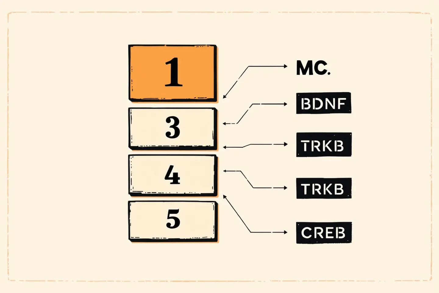
Semax is a synthetic analog of ACTH(4-10). Like its parent fragment, it appears to engage the melanocortin receptor family — the literature describes partial agonism, antagonism, or mixed activity at MC3R, MC4R, and MC5R, and the precise pharmacology has not been definitively resolved. What is well-replicated is the downstream effect: in rat brain tissue, Semax upregulates BDNF and NGF and modulates the dopaminergic and serotonergic systems.
Dolotov and colleagues established the canonical mechanism finding. A single intranasal dose of 50 mcg/kg in male Wistar rats produced a 1.4-fold increase in hippocampal BDNF protein, a 1.6-fold increase in TrkB tyrosine phosphorylation, an approximately three-fold increase in exon III BDNF mRNA, and a doubling of trkB mRNA, alongside improved performance on a conditioned avoidance task [1]. The companion binding study identified specific, reversible binding of Semax to rat basal forebrain membranes with a dissociation constant of 2.4 ± 1.0 nM and demonstrated that the resulting BDNF upregulation was regionally selective — present in basal forebrain at three hours, absent in cerebellum [2].
A later time-course study added nuance. Across rat hippocampus, frontal cortex, and retina, Semax produced multidirectional, tissue-specific regulation of NGF and BDNF mRNA: brief decreases in hippocampus and retina at 20 minutes post-dosing, increases in frontal cortex, with retinal BDNF significantly elevated at 90 minutes [8]. The signal is not a uniform upregulation; it is a regional and temporal program.
In vitro, Semax inhibits enkephalin-degrading enzymes of human serum with an IC50 near 10 μM — more potent inhibition than puromycin or bacitracin under comparable conditions. The activity localizes to the pentapeptide fragments of Semax and the related Russian peptide Selank; shorter (three- to four-residue) and six-residue fragments are inactive [9]. This pathway is hypothesized to prolong endogenous opioid signaling and may contribute to the analgesic effects reported in rodent compression assays [16].
The 2022 ACS Chemical Neuroscience paper added a copper-chelation mechanism: Semax demonstrates high affinity for Cu(II) ions and produces concentration-dependent inhibition of copper-induced amyloid-beta aggregation in cell-free and artificial membrane models, with approximately 90% reduction in amyloid-beta cytotoxicity in differentiated SH-SY5Y neuroblastoma cells [12]. The 2025 Bulletin of Experimental Biology and Medicine paper added direct modulation of intracellular Ca2+ dynamics in rat brain neurons, suggesting ion-handling effects upstream of the BDNF/CREB transcriptional program [17].
SECTION 02 / PRECLINICAL ISCHEMIA
The rat middle cerebral artery occlusion model is the most-studied preclinical context for Semax. Sudarkina and colleagues, working in the Myasoedov group, documented the protein-expression signature after transient MCAO and 100 mcg/kg intraperitoneal Semax: active CREB increased more than 1.5-fold in subcortical ischemic structures (a pro-survival transcription factor activation), active JNK decreased more than 1.5-fold across cortex and subcortex (suppression of apoptotic and inflammatory signaling), and MMP-9 expression decreased approximately 1.5-fold in adjacent frontoparietal cortex (reduction of a blood-brain-barrier-disrupting protease) [4].
In permanent MCAO, Dmitrieva and colleagues compared intact Semax (100 mcg/kg IP) against the active metabolite Pro-Gly-Pro (37.5 mcg/kg IP) and found that both activated expression of neurotrophin and neurotrophin-receptor genes (BDNF, NGF, and their receptors) in damaged cortex at 3, 24, and 72 hours after injury. Semax demonstrated greater spatial selectivity to the injured tissue than PGP administered alone [5].
Medvedeva and colleagues extended this with genome-wide RNA-Seq after transient MCAO. The 394 differentially expressed genes resolved into a coherent program: suppression of inflammatory and immune-system transcripts paired with activation of neurotransmitter-system genes, with the largest effect window between 3 and 24 hours after the ischemic insult — the same window during which penumbral cells remain salvageable [6]. Romanova and colleagues reported the functional correlate: 50 mcg/kg intranasal Semax for six days reduced cortical infarct volume after focal photoinduced ischemia and preserved retention of conditioned passive avoidance [7].
SECTION 03 / RUSSIAN CLINICAL EVIDENCE
The Russian clinical record on Semax begins with the Gusev group's 1997 open-label study in 30 patients with acute hemispheric ischemic stroke. Patients received intranasal Semax 1% — 12 mg/day for moderate strokes (five-day course) or 18 mg/day for severe strokes (ten-day course) — alongside conventional therapy. Compared to 80 control patients on conventional therapy alone, the Semax arm showed accelerated regression of general cerebral and focal motor deficits, with parallel improvements on EEG mapping and somatosensory evoked potentials [3]. The paper is published in Zhurnal Nevrologii i Psikhiatrii imeni S.S. Korsakova in Russian; the English-language record consists of the PubMed abstract.
The recovery-phase study (Gusev, Martynov, and colleagues) followed 110 post-stroke patients (43 men, 67 women, mean age 58.0 ± 9.7 years) receiving 6,000 mcg/day intranasal Semax in two ten-day courses separated by a twenty-day interval. The cohort showed elevated plasma BDNF and improvements on the Barthel index of activities of daily living and the MRC motor scale [14]. This citation is reconstructed from secondary sources; the primary Russian-language publication was not independently verified in PubMed at the time of writing.
The ophthalmology literature carries the second documented human indication. Polunin and colleagues reported on 36 patients (72 eyes) with primary open-angle glaucoma and normalized intraocular pressure who received Semax 0.1% intranasal as an adjunct to standard topical neuroprotective therapy. The Semax adjunct arm showed electrophysiologic and computer-perimetry improvements over conventional treatment alone, with no negative changes in optic nerve head structure or corneal subbasal nerve plexus during the observation period [15]. The paper is published in Vestnik Oftalmologii in Russian.
No large, multi-center, double-blind randomized controlled trial of Semax meeting current Western regulatory standards has been published in a major Western journal. As a result, the FDA, EMA, MHRA, and Health Canada have not evaluated the compound, and the strength of evidence relative to Western pharmaceutical standards remains limited.

SECTION 04 / RECENT WESTERN-JOURNAL WORK
Three Western-journal papers between 2022 and 2025 mark a quiet expansion of the mechanism literature. Inozemtseva and colleagues, in a 2024 European Journal of Pharmacology paper, applied a chronic unpredictable stress (CUS) rat model of depression and found that daily intraperitoneal Semax at 60 nmol/kg reversed or substantially attenuated stress-induced anhedonia (sucrose preference), suppressed body-weight gain, adrenal hypertrophy, and the decrease in hippocampal BDNF. The non-selective melanocortin analog Melanotan II at the same low dose produced comparable antidepressant-like effects, supporting the melanocortin-system framing of the mechanism [10].
Liu and colleagues, in a 2025 British Journal of Pharmacology paper, reported a novel axis in a thoracic (T9–T10) spinal cord injury model in female C57BL/6 mice. Semax improved functional recovery and inhibited lysosomal-membrane-permeabilization-driven pyroptosis. RNA-seq and knockdown experiments mapped the mechanism to mu-opioid receptor (Oprm1) signaling regulating USP18, which deubiquitinates the fat-mass-and-obesity-associated protein (FTO) [11]. This is the first published Semax mechanism that extends beyond the classical melanocortin and BDNF pathways.
Sciacca and colleagues, in the 2022 ACS Chemical Neuroscience paper noted in the mechanism section, provided the copper-chelation evidence [12]. The 2025 Bulletin of Experimental Biology and Medicine report on intracellular calcium dynamics in rat brain neurons added the ion-handling layer [17]. Together these four papers represent a slow but real broadening of the Semax mechanism record outside the original Russian Academy of Sciences program.